MEG Overview

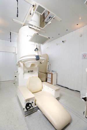
Magnetoencephalography (MEG) is a non-invasive procedure similar to electroencephalography (EEG) in terms of basic principles and analysis, however, MEG consists of sitting in a chair or lying on a bed while your head is inside a helmet shaped device which contains magnetic field sensors. These superconducting quantum interference device (SQUID) sensors passively detect the weak magnetic fields (around 10-14 Tesla) produced by brain activity. In the NIH scanner, 275 SQUID sensors are uniformly distributed, in a grid, over the inner surface of a helmet that covers the entire head. Head position within the magnetometer is determined before and after the MEG session by digitizing the position of three indicator coils that are attached to the pre-auricular and the nasion fiducial points. These positions define the coordinate system for the signals.
Magnetoencephalography can lead to a better understanding of the functioning of the brain because the potential to determine the spatial source of the recorded activity is enhanced. With this technology signal detection is practically unaffected by the conductivity and structure of skull and scalp tissue. In addition, compared to EEG systems, MEG systems allow higher spatial sampling resolution. Under favorable conditions, spatial localization of current sources with whole head MEG is on the order of 2–3 mm at a temporal resolution better than 1 ms.
For more information, the wikipedia page on MEG is useful.
There are also several introductory books of interest: MEG: An Introduction to Methods, Magnetoencephalography: From Signals to Dynamic Cortical Networks, and MEG-EEG Primer.